Mitral regurgitation
LV → LA
Backward flow across the mitral valve while the LV contracts. In the same injection, LV → aorta is expected forward ejection.
Into the left atrium. During the squeeze, watch for a second chamber filling with contrast beside the LV.
Real published cineRAO 30° verified in the sourceOptional labelsThe clinical video is unchanged. Colored pointers are a separate teaching layer; they mark approximate regions, not traced chamber borders.
Konishi et al., 2016 · Video S3 · 6.73 seconds. Figure 3 confirms RAO 30°. The authors report grade 3–4 MR.
The clue may be a spreading gray cloud in the LA. Ask: “As the LV squeezes, does a second chamber fill?”
LA contrast can remain visible after systole; watch when it first enters or increases.
“Left” here means screen-left in this example, not the patient’s anatomical left. Use the actual projection and landmarks on each new cine.
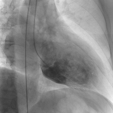
Find the large LV on screen-right and its pigtail catheter.
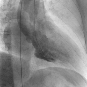
LV cavity narrows; watch the left atrial region beside the spine.
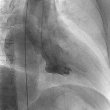
The second, screen-left contrast-filled chamber is the key.
Frames extracted from the original cine without altered anatomy, contrast enhancement, or generated pixels. Phase descriptions are based on visible cavity change; there is no synchronized ECG in this clip.
The LV projects down and to the right. The LA is behind the LV anatomically and projects toward screen-left, near the spine. The aortic outflow rises above the LV.
First find the LV and watch it contract. Then look outside its basal border for contrast entering the LA.
Do not call every plume above the LV “MR.” Identify which chamber or vessel receives it, and when.
Projection and overlap matter. The aorta, atrium, spine, and catheter may overlap; a fixed screen direction alone is not diagnostic.
Backward flow across the mitral valve while the LV contracts. In the same injection, LV → aorta is expected forward ejection.
Contrast leaks back through the aortic valve into the LV during relaxation. An ordinary LV injection already fills the LV, so aortic opacification from that injection does not establish AI.
After an LV injection, contrast enters the chamber beside the spine as the LV becomes smaller.
This is a source-based educational example, not recalled examination content. It does not establish which projection or image any examination will use.
Back to unlabelled practice →